
Why Brain Function May Be the Earliest Signal of Brain Health
From aging to injury, functional signals captured through MEG can often appear before visible structural damage.
When most people think about getting a brain scan, they picture something like an MRI. The images are powerful and detailed. They can show tumours, bleeding, or structural damage. But many conditions, especially those linked to memory and cognition, don’t start with something you can see. They begin with something you can only measure: brain function.
At MYndspan, we use magnetoencephalography (MEG) because it shows us how the brain is really functioning in real time. MEG has long been used in neurology to plan brain surgeries or pinpoint where epileptic seizures begin. It’s non-invasive, silent, and radiation-free, capturing neural activity with millisecond precision.
But its potential goes far beyond epilepsy. For anyone looking to understand their cognitive health or detect early decline, function might be the first and most important signal.
Form vs. Function: What Are We Actually Measuring?
MRI tells us about structure—how the brain looks. It identifies tumours, lesions, bleeding, or areas of shrinkage. Functional MRI (fMRI) shows blood flow patterns related to brain activity. These are essential tools, especially when structural abnormalities are involved.
MEG, on the other hand, captures the magnetic fields produced by neurons as they fire. It measures neural activity directly, not through blood flow, and does so with millisecond accuracy.
As the Cleveland Clinic explains:
“MEG is “the most advanced method of recording and evaluating your brain activity… [It] maps out the sensory areas of your brain and can pinpoint the exact location where seizures originate.”
Importantly, MEG isn’t only for detecting seizures or planning surgeries. It’s increasingly being used to study how the brain changes in aging, injury, and early neurodegeneration, often before MRI reveals anything unusual.
A study published in Human Brain Mapping compared MEG and fMRI during a picture-naming task. Both methods showed similar overall patterns of brain activity, but MEG revealed details that fMRI missed.
That’s because the two techniques work very differently:
MEG captures synchronised neural activity with millisecond precision.
fMRI measures slower changes in blood flow, averaged over time.
This difference is crucial. In the study, MEG detected rapid activity in the visual cortex that fMRI couldn’t pick up, because the stimulus was too brief to register in the slower blood-flow signal.
In practice, this means MEG can identify subtle changes in brain function, such as slowing, disrupted connectivity, or unusual network behaviour, well before structural changes appear. These are often the earliest signs of conditions like concussion effects or neurodegenerative disease.

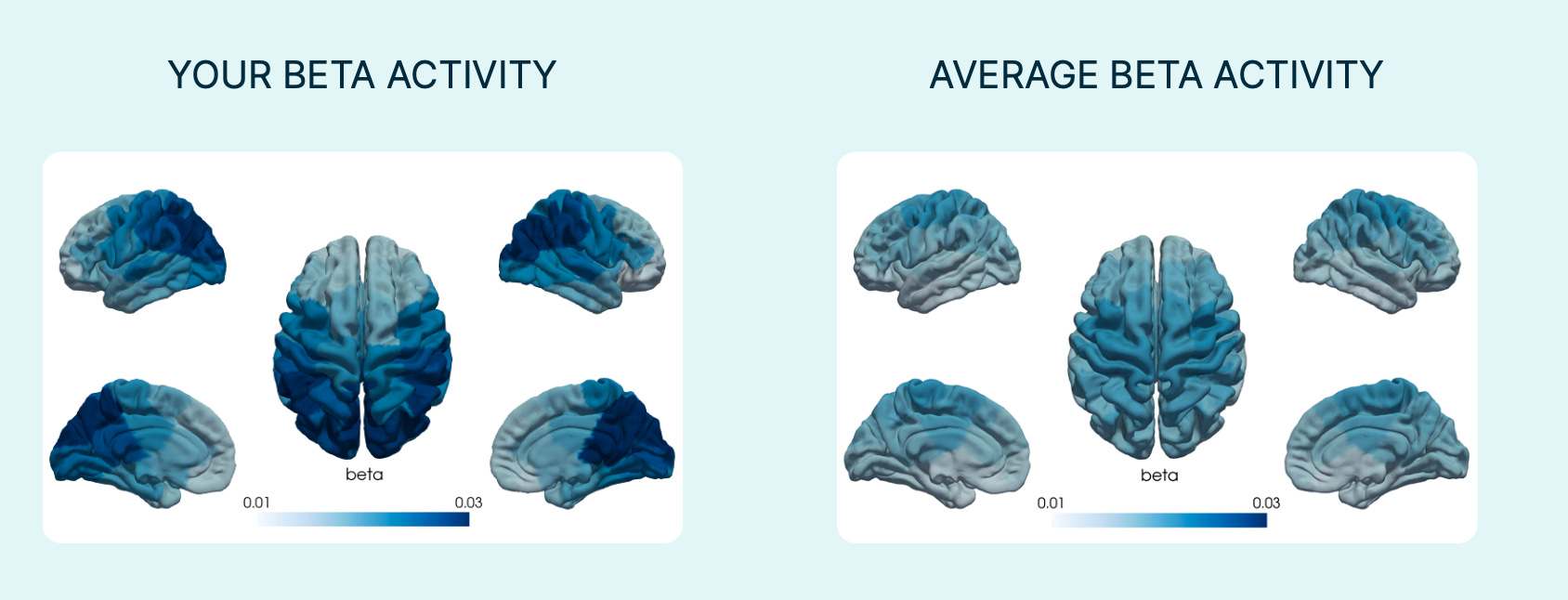
The images above show what you’d typically see from PET, MRI, and fMRI scans — focused on brain structure and blood flow. In contrast, MYndspan’s MEG-based maps (below) highlight a snapshot of brainwave activity — here comparing a client’s beta activity to the population average.
MEG and the Promise of Early Detection
Neurodegenerative diseases like Alzheimer’s disease and other dementias don’t appear overnight. They begin as functional disruptions, or changes in how the brain communicates, processes information, or maintains network stability.
MEG is uniquely equipped to detect these early signs. Studies have shown its sensitivity to:
Accelerated brain ageing
Dysconnectivity in brain networks
Changes in brainwave timing and synchrony
These patterns often appear before structural atrophy, which is what MRI is designed to capture.
At MYndspan, we use MEG to assess:
Functional Brain Age: a measure of how fast or slow your brain is operating relative to your chronological age
Brain Stability Index: a measure of how stable brain connectivity is over time. Shifts in this stability can reveal early signs of stress, injuries like concussion or cognitive decline.
Brainwave Mapping: patterns related to focus, sleep, and stress response
All of this is captured in just a 10-minute MEG scan - completely safe, comfortable, and free from radiation and injections.
MEG Doesn’t Replace MRI, It Complements It
MRI is essential. It provides structural detail and can detect abnormalities like tumours, haemorrhages, or brain shrinkage. But structure doesn’t tell the whole story.
As we often say:
“MRI tells you what your brain looks like. MEG tells you what it’s doing.”
They complement each other. MEG adds a layer of information that helps fill in the picture, especially when looking for early or subtle functional changes. Preventive brain health isn’t just about reacting to symptoms. It’s about identifying patterns, like slowing, decline, or network disruptions, before symptoms appear. By detecting these functional changes early, MEG makes it possible to intervene sooner, when treatments and lifestyle changes are most effective.
As research advances, and as we continue to bring this technology out of academic labs and into public spaces, brain function will become a standard part of how we assess and protect brain health.
At MYndspan, our mission is to end preventable neurodegeneration through early detection. MEG is a critical part of that future.