


Why MEG is the Gold Standard for Measuring Brain Function
Magnetoencephalography (MEG) is a non-invasive imaging technology that has been used for decades in clinical and research settings. We're unlocking its value for more widespread adoption.

We have had an exciting few months here at MYndspan and have a considerable amount of new followers and subscribers – welcome!
We tend to receive quite a few questions about the technology we use – magnetoencephalography (it’s a mouthful, so we just say MEG).
Our focus today is a deep-dive on all things MEG and to answer as many questions as we can. But we don’t want it to be a one way conversation! Please never hesitate to reach out and ask us questions or let us know if you want us to cover a specific topic as it relates to proactive and preventive brain health.
Magnetoencephalography (MEG) is a non-invasive neuroimaging technique used to measure the magnetic fields produced by neurons in the brain. It provides both high temporal and spatial resolution, making it valuable for both research and clinical applications.
Definition time:
High temporal resolution means that MEG provides high accuracy when it comes to the timing of brain activity measured (to the millisecond).
High spatial resolution means that MEG provides high accuracy when it comes to the location of where that activity is coming from (to the millimeter).
Surprisingly, MEG isn’t a new technology. MEG scanners have actually been used for decades to measure brain function and activity in clinical and research settings.
But we believe that this incredible technology has a much bigger role to play in brain function and health analysis. We are steadfastly focused on unlocking MEG for all people interested in taking a more proactive and preventive approach to their overall brain health.
Book your scan or join the waitlist for our other locations here.
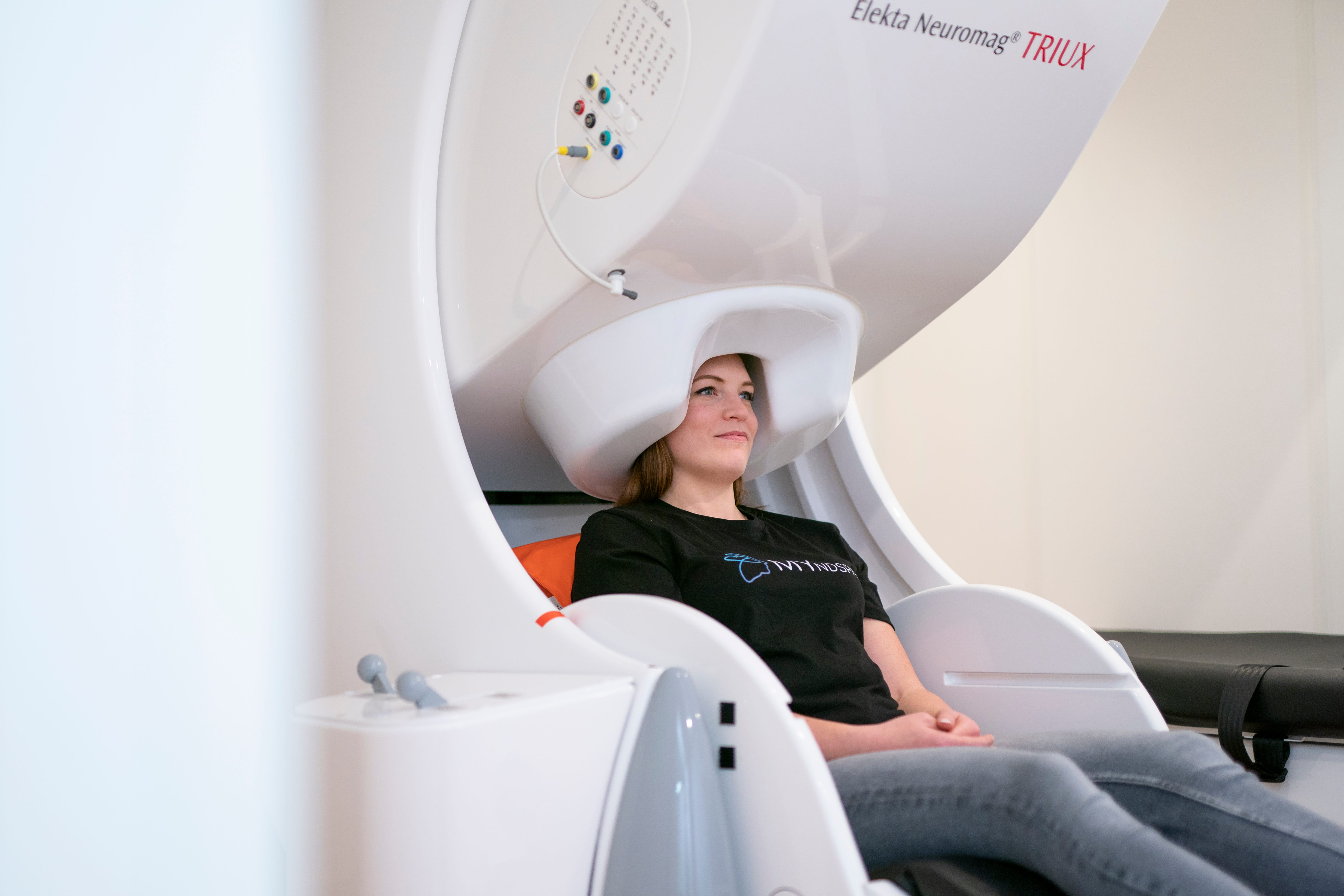
What does MEG do?
Above is an informative video from MEGIN, the global leaders in manufacturing MEG devices, who describe how the scanner works, and how it is used.
MEG scanners detect and record the activity of your brain, differing from other well-known neuroimaging modalities such as magnetic resonance imaging (MRI), which takes snapshots of the physical structure of your brain.
Whereas the output of an MRI shows an individual their anatomy, MEG is focused on neurophysiology, or the functioning of your brain. In this way, these two technologies serve slightly different purposes, but they can complement each other and are often used together.
When your neurons communicate, they produce electrical currents that flow through the brain. These currents generate weak magnetic fields, which MEG sensors can detect. MEG offers the best temporal resolution which allows for the precise timing of neural events.
With its accuracy in measuring brain function (rather than structure like other modalities), MEG technology has the potential to identify issues before symptoms even emerge.

Who uses MEG today?
MEG has been used for decades across both clinical and research settings.
Clinical: MEG is used in the diagnosis and pre-surgical planning for epilepsy, as it can accurately localize the regions of the brain where seizures originate. It is also used for mapping brain function before brain surgery to avoid critical areas.
Research: MEG is widely used in cognitive neuroscience to study sensory processing, language, memory, and in clinical research for both neurodegeneration (like Alzheimer’s) and neuropsychiatric disorders (like PTSD). It helps researchers understand how different regions of the brain interact in real-time.
Where is MEG going?
MEG for disease detection
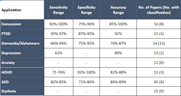
A number of papers exist which use data from MEG scans to automatically identify subjects with or without a given health issue.
MEG offers a new way to assist in disease detection, alleviating the need to solely rely on self-reported or semi-subjective symptoms, and instead allowing for inspection of the root cause. For example, MEG has provided new insights into functional changes and disease progression for neurodegenerative diseases including Alzheimer’s and Parkinson’s disease.
Why is this important? Well, much of our understanding of mental phenomena is based on long-held beliefs derived from psychology, which are not always aligned with biological reality.
MEG has also proven to be useful in studying and diagnosing neurodevelopmental disorders such as autism spectrum disorder (ASD) and attention deficit hyperactivity disorder (ADHD) by identifying atypical patterns of brain activity.
MEG for concussion recovery
MEG has a promising role to play in the assessment and management of concussions. It can detect subtle functional abnormalities in brain activity that may not be visible through other imaging techniques like CT or standard MRI, and can identify disruptions in neural networks, helping to understand how a concussion has affected brain connectivity and communication. A concussion is an invisible functional injury that MEG can measure, especially when one has a baseline measure to compare to.
MEG for longevity and proactive health
MEG technology offers significant potential for advancing our understanding of brain aging and promoting longevity. By providing detailed insights into brain function, monitoring changes over time, evaluating interventions, and guiding personalized strategies, MEG can play a crucial role in helping individuals maintain cognitive health and enhance their quality of life as they age.
Why MEG?
In our next post, we are going to look at how MEG compares to other brain assessment tools, including fMRI and EEG. There are specific use-cases and situations where each technology shines, and they often work together. When it comes to MEG, a few things stand out:
Non-invasive and safe, with no exposure to ionizing radiation like you would require for a PET scan.
High temporal resolution, allowing for the tracking of rapid neural activity - scanning at the speed of thought.
High spatial resolution, providing a detailed map of brain activity and its location.
Easily tolerated, no noises or confining spaces like that of MRI.
Beyond these, one of the things that excites us most in MEG’s potential lies in its repeatability.
MEG’s safety profile allows for repeated measurements over time, which is essential for longitudinal studies and monitoring disease progression or treatment effects.
You can have a scan as often as you’d like. Because it is a passive recording device, there are no negative side effects. By receiving routine assessments over time, you can establish a baseline of healthy brain connectivity and detect any deviations from the norm.
Why hasn't MEG been used more?
MEG scanners are not as ubiquitous as MRI scanners or EEG devices, so one limitation is direct access. However, there are enough scanners to unlock its potential as a preventive brain health modality across all of the above use cases.
The main impediment to using it at a larger, non-research scale is the complexity required when it comes to analyzing MEG data, and the very specific skill needed to do so.
This is where we come in.
Book your scan or join the waitlist for our other locations here.